
What “brain mapping” really means?
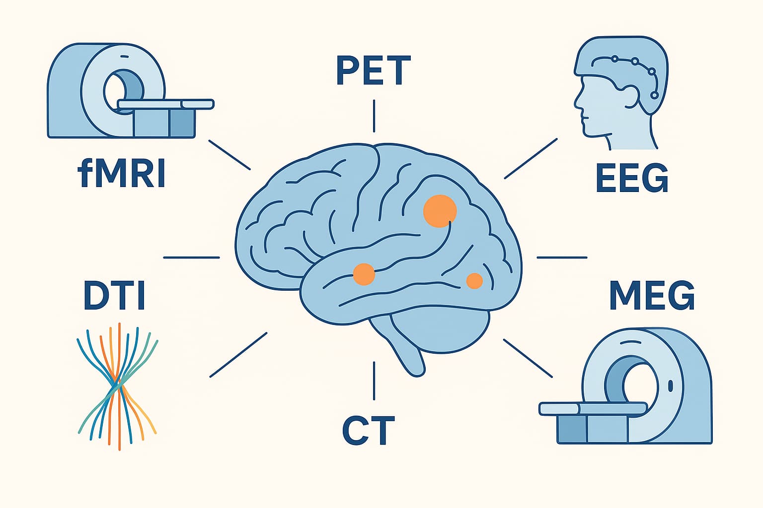
Brain mapping is the toolbox of methods used to chart the brain’s structure (what it looks like and how it’s wired), function (what areas are active and when), and connectivity (how regions interact). In practice, labs and clinics combine multiple techniques to trade off spatial detail, timing precision, cost, and invasiveness.
Structural vs functional mapping (and connectivity)
-
-
Structural mapping (anatomy): CT, structural MRI, and diffusion MRI depict tissue types and white-matter pathways. Think “parts and wiring.”
-
Functional mapping (activity): fMRI, PET, EEG, and MEG capture physiology linked to neural activity. Think “when and where it fires.”
-
Connectivity mapping:
-
Structural connectivity: diffusion MRI tractography infers axonal pathways.
-
Functional connectivity: temporal correlations in fMRI, EEG, or MEG signals (which regions fluctuate together).
-
Effective connectivity: models directed influence (e.g., dynamic causal modeling, Granger causality).
-
-
Core techniques at a glance
| Modality | What it measures | Spatial resolution | Temporal resolution | Invasiveness | Typical uses |
|---|---|---|---|---|---|
| CT | X-ray attenuation (tissue density) | ~0.5–1 mm | Seconds | Non-invasive, ionizing radiation | Acute hemorrhage, fractures, mass effect, CT perfusion in stroke |
| Structural MRI (T1/T2) | Proton relaxation (tissue contrast) | ~0.5–1 mm at 3T; sub-mm at 7T | Seconds–minutes (per volume) | Non-invasive | Anatomy, volumetry, lesion load, cortical thickness |
| DTI / diffusion MRI | Directional water diffusion (microstructure) | 1–2 mm voxels (clinical); sub-mm research | Minutes (per scan) | Non-invasive | White-matter tracts, structural connectivity, pre-surgical tractography |
| fMRI (BOLD, ASL) | Hemodynamics (blood oxygenation/flow) as proxy for neural activity | 2–3 mm typical; ≤1 mm at 7T | ~1–2 s TR; neural events blurred (~5–7 s hemodynamics) | Non-invasive | Cognitive mapping, resting-state networks, presurgical localization |
| PET | Radiotracer uptake (metabolism or specific targets) | 3–5 mm (modern PET/CT or PET/MR) | Tens of seconds–minutes | Minimally invasive (radiotracer, ionizing radiation) | Oncology, neurodegeneration (amyloid/tau), epilepsy, neuroinflammation |
| EEG | Scalp electrical potentials from synchronous post-synaptic activity | Centimeters on cortex after source modeling | Milliseconds | Non-invasive | Epilepsy, sleep, evoked potentials, neurofeedback, BCI |
| MEG | Magnetic fields from cortical currents | ~3–5 mm with source modeling | Milliseconds | Non-invasive | Epilepsy source localization, sensory/motor timing, network dynamics |
Notes:
-
ASL fMRI quantifies cerebral blood flow. BOLD fMRI is the most common.
-
PET tracers can target glucose metabolism (FDG), β-amyloid, tau, dopamine, or synaptic density (SV2A), among others.
-
EEG/MEG have superb timing but require source modeling to localize activity.
-
Invasive options (not the main focus here) include ECoG and depth electrodes for ultra-precise clinical mapping in refractory epilepsy.
How the major techniques work
CT
-
Physics: Rotating X-ray source + detectors reconstruct tissue density.
-
Strengths: Speed, availability, excellent for blood, bone, and emergencies.
-
Limits: Radiation exposure; poorer soft-tissue contrast vs MRI.
Structural MRI (T1/T2/FLAIR)
-
Physics: Aligns proton spins in a magnetic field; radiofrequency pulses probe relaxation properties.
-
Strengths: Superb soft-tissue contrast, lesion detection, volumetry, myelin-sensitive contrasts.
-
Limits: Longer scans, motion sensitivity, contraindications (some implants).
DTI / diffusion MRI
-
Physics: Diffusion-sensitizing gradients quantify water motion; tensors or advanced models (NODDI, DKI) infer microstructure.
-
Strengths: Models anisotropy to reconstruct white-matter tracts.
-
Limits: Crossing fibers and partial-volume effects can mislead tractography; indirect inference.
fMRI (BOLD and ASL)
-
Physics: Neural activity alters local blood oxygenation/flow; MRI detects the hemodynamic signature.
-
Strengths: Whole-brain coverage, good spatial resolution, rich connectivity analyses (task and resting state).
-
Limits: Indirect measure (neurovascular coupling), seconds-scale delay, susceptibility to motion/physiology.
PET
-
Physics: Injected radiotracers emit positrons; annihilation photons detected in coincidence reconstruct uptake maps.
-
Strengths: Molecular specificity (metabolism, receptors, aggregates), disease staging, treatment monitoring.
-
Limits: Radiation, cost, limited temporal sampling; requires cyclotron/radiochemistry for some tracers.
EEG
-
Physics: Scalp electrodes measure summed cortical postsynaptic potentials.
-
Strengths: Millisecond timing, portable, low cost.
-
Limits: Blurred spatial localization due to skull/scalp; the electromagnetic inverse problem is ill-posed.
MEG
-
Physics: SQUIDs or optically pumped magnetometers sense femto-tesla magnetic fields from neuronal currents.
-
Strengths: Millisecond timing with better localization than EEG (less skull distortion).
-
Limits: Expensive, magnetically shielded rooms (for SQUID systems), mainly sensitive to tangential cortical sources.
What “resolution” realistically looks like
-
Spatial: MRI/DTI/fMRI resolve millimeter-scale voxels; PET ~3–5 mm; EEG/MEG source maps are typically centimeters unless constrained with MRI.
-
Temporal: EEG/MEG capture millisecond dynamics; fMRI captures slow hemodynamics (~1–2 s sampling with several-second lag); PET captures minute-scale trends.
Clinical applications
Pre-surgical planning
-
Tumors/lesions: Structural MRI delineates margins; DTI maps tracts (e.g., corticospinal, arcuate) to minimize deficits; task fMRI localizes language/motor areas; MEG/EEG or ECoG refine eloquent cortex.
-
Epilepsy: EEG/MEG localize interictal spikes; PET (interictal hypometabolism) and ictal SPECT; MRI finds focal cortical dysplasia; invasive monitoring when needed.
Acute and chronic disorders
-
Stroke: CT for hemorrhage; CT/MR perfusion and diffusion to determine salvageable penumbra; follow-up MRI for recovery mapping.
-
Neurodegeneration: PET amyloid/tau for Alzheimer’s characterization; structural MRI for atrophy patterns; FDG-PET for hypometabolism.
-
Movement disorders: MRI/DWI for surgical planning (DBS targets); MEG/EEG for oscillatory biomarkers.
-
Psychiatry (emerging): PET for inflammation/receptor status in research; fMRI connectivity phenotypes to subtype depression or predict treatment response (research to clinic pipeline).
Treatment monitoring
-
Oncology: Serial MRI/PET to track tumor response.
-
Rehabilitation: fMRI/EEG biomarkers of reorganization after stroke; connectivity changes as proxies for recovery.
-
Neurofeedback: Real-time fMRI or EEG trains patients to modulate activity in targeted circuits (e.g., pain, mood, attention).
Research applications
Cognitive and systems neuroscience
-
Task fMRI: Map functional specialization (visual, language, memory).
-
Resting-state fMRI: Intrinsic networks (default mode, salience, frontoparietal control).
-
EEG/MEG: Temporal sequencing of perception/action; oscillations (theta, alpha, beta, gamma) and cross-frequency coupling.
Connectivity and network science
-
Structural connectomes: Diffusion tractography at individual and population scales.
-
Functional connectomes: Correlation and graph-theoretic metrics (degree, modularity, hubs).
-
Effective connectivity: Directional models to test causal hypotheses.
Population studies and precision neuroscience
-
Large cohorts: Harmonized protocols uncover variability across age, sex, and disease.
-
Individual-level mapping: Subject-specific parcellations improve reproducibility and clinical translation.
Recent and notable developments
Higher field and higher resolution MRI
-
7-Tesla MRI: Sub-millimeter cortical layer-specific fMRI, finer subcortical nuclei, better susceptibility contrast.
-
Advanced sequences: Multi-band EPI for faster fMRI; vascular-space-occupancy and calibrated fMRI to separate blood flow vs oxygen metabolism; ASL for quantitative perfusion.
Better diffusion models and tractography
-
Beyond tensors: Multi-shell acquisitions (e.g., CSD, NODDI) handle crossing fibers; microstructural indices (neurite density, orientation dispersion) improve biological specificity.
PET innovation
-
New tracers: Tau, synaptic density (SV2A), neuroinflammation (TSPO alternatives), α-synuclein in development.
-
Total-body PET: Higher sensitivity enables lower dose and dynamic whole-axis imaging.
Fast and wearable neurophysiology
-
High-density EEG with improved source modeling and artifact correction.
-
OPM-MEG (wearable): Moves with the head, enabling studies in more natural settings and pediatric populations.
Real-time and closed-loop mapping
-
Real-time fMRI (rt-fMRI): Neurofeedback and adaptive paradigms.
-
Closed-loop EEG/MEG: Stimulate contingent on ongoing brain state (TMS-EEG, tACS-EEG) to probe causality.
Multimodal integration and hybrid systems
-
Simultaneous EEG-fMRI to link millisecond timing with millimeter localization.
-
Hybrid PET/MR for concurrent molecular and structural/functional data.
-
Data fusion/ML: Deep learning for segmentation, denoising, artifact removal, and multimodal feature integration; normative modeling to detect patient-specific deviations.
Limitations and challenges
Biological and methodological caveats
-
Indirect signals: fMRI reflects hemodynamics, not spikes; neurovascular coupling varies with age/disease/drugs.
-
Inverse problem: EEG/MEG localization is underdetermined; accuracy depends on head models and priors.
-
Tractography pitfalls: Crossing/branching fibers, gyral biases, and false positives/negatives.
-
PET specificity: Off-target binding, tracer kinetics, and partial-volume effects complicate interpretation.
Practical constraints
-
Cost and access: PET/MR and MEG are expensive; CT and MRI are widely available but still resource-intensive.
-
Motion and artifacts: Pediatric, movement-disorder, or critically ill patients can be hard to scan.
-
Interpretation complexity: High-dimensional data require careful statistics, preregistration, and replication to avoid false findings.
Ethics and data governance
-
Incidental findings: Require clinical review pathways.
-
Privacy: Brain data are sensitive; de-identification, consent, and secure storage are essential.
-
Equity: Ensure diverse populations in datasets to avoid biased tools and conclusions.
Putting it together in practice
Typical clinical pipeline (example: brain tumor)
-
Structural MRI for anatomy and edema.
-
DTI tractography to map eloquent tracts (e.g., language, motor).
-
Task or resting-state fMRI to localize functional regions.
-
MEG/EEG if seizures or precise timing questions arise.
-
Intraoperative mapping (when indicated) to preserve function.
Typical research pipeline (example: language network)
-
High-res structural MRI for cortical surfaces and parcellation.
-
Task fMRI (semantic/syntactic contrasts) + resting fMRI for connectivity.
-
EEG/MEG to resolve timing of lexical vs syntactic processing.
-
Diffusion MRI to relate function to white-matter pathways (arcuate, SLF).
-
Multimodal modeling (e.g., representational similarity, ML decoding).
Choosing the right tool: quick guide
-
Need millisecond timing? EEG/MEG.
-
Need millimeter localization across the whole brain? fMRI.
-
Need white-matter wiring? Diffusion MRI (DTI/CSD).
-
Need molecular information (amyloid, metabolism, receptors)? PET.
-
Need speed in emergencies or bone/blood detection? CT.
-
Need high-contrast anatomy and volumetry? Structural MRI.
Key takeaways
-
No single modality is “best.” Each observes a different facet of the brain.
-
Multimodal strategies are the norm: combine who/where (MRI/DTI), when (EEG/MEG), and what biology (PET).
-
Advances in ultra-high-field MRI, OPM-MEG, total-body PET, and AI-driven multimodal fusion are pushing resolution, speed, and interpretability.
-
Clinical impact is strongest in surgical planning, stroke, epilepsy, oncology, and neurodegeneration, with growing roles in psychiatry and rehabilitation.
-
Rigorous methods, transparent reporting, and careful inference are essential to translate brain maps into reliable science and effective care.
